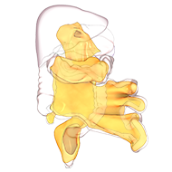
Section 6.4 – Lateral mobilisation of the mesosigmoid and left mesocolon
Left sided mobilisation of the mesentery begins by retracting the mesosigmoid medially
This exposes congenital adhesions that can be divided
These are highly variable but always present
They should not be mistaken with regions of the reflection
With which they can fuse
Their division exposes the reflection
When the reflection is divided through
The undersurface of the mesosigmoid is apparent
The mesosigmoid is detached,
But maintained fully intact at all points
The mesosigmoid is continuous with the left mesocolon
Once the mesosigmoid has been detached,
The in continuity left mesocolon becomes apparent
This is also detached, in the same manner, from the underlying fascia
This way, it is maintained intact, during the detachment process
The key to detachment of an intact mesentery
Is to identify the zone (or plane) between mesentery above and fascia below
This plane is always present, and is the roadmap used in surgical dissection
The plane is demonstrated, by lifting the mesentery away from the fascia
Thereby placing the interface between fascia and mesentery under stretch
Separation of the mesentery from fascia, is called mesofascial separation
It can be achieved by sharp or blunt dissection
But always requires a clear field of view
And unimpeded access
With continued mesofascial separation, the undersurface of the mesosigmoid becomes apparent
