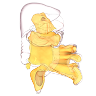
Section 6.17 Attachment: The fascia and reflection
The fascia, between mesenteric and non-mesenteric domains
Is thin and very flimsy in areas
As a result, it is readily disrupted during dissection
Notwithstanding this
Its anatomical distribution is important and can be demonstrated
The thickness of the fascia varies between regions
For example, that under the dorsal mesogastrium is dense and easily demonstrated
The distribution of the divided edge of the reflection can also be determined
Here we are seeing the divided edge of the hepatocolic, right lateral and ileocolic regions of the reflection
This is where the parietal peritoneum is reflected onto the mesenteric domain
The divided edge of the reflection can also be seen on the left
In fact it can be followed from the DJ flexure
Medially down into the pelvis,
Then back up around the left
Under the diaphragm,
Down the right side, and medially back up to the
DJ flexure
Certain regions are best visualised after removal of the mesenteric domain
These include the lienophrenic and coronary regions
